Ihd Dilatation Ct

Precontrast Ct Shows Ihd And Cbd Dilatation With Several Radiopaque Download Scientific Diagram

Precontrast Ct Shows Ihd And Cbd Dilatation With Several Radiopaque Download Scientific Diagram

Intrahepatic Duct Dilatation Unknown Cause Radiology Case Radiopaedia Org

Intrahepatic Duct Dilatation Unknown Cause Radiology Case Radiopaedia Org

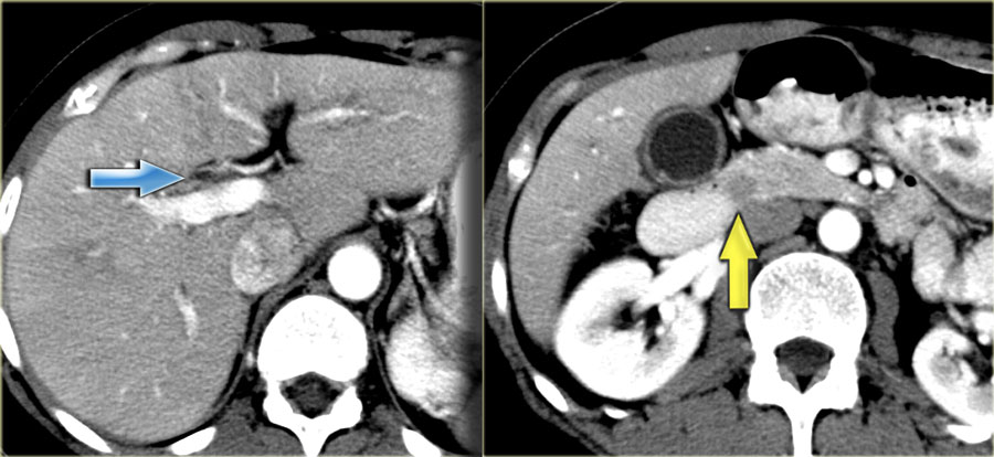
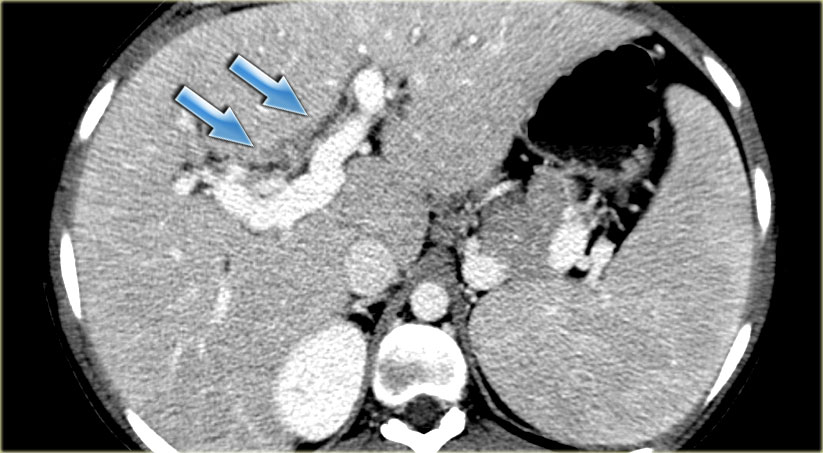
The Radiology Assistant Biliary Duct Pathology

The Radiology Assistant Biliary Duct Pathology

The Radiology Assistant Biliary Duct Pathology

The Radiology Assistant Biliary Duct Pathology

Contrast Enhanced Computed Tomography Scan Finding It Shows The Download Scientific Diagram

Contrast Enhanced Computed Tomography Scan Finding It Shows The Download Scientific Diagram

Https Car Ca Uploads Education 20lifelong 20learning Meetings Asm2015 Speakers Pres Ee014 Approach To Imaging Of The Dilated Bile Duct Zhang Pdf

Https Car Ca Uploads Education 20lifelong 20learning Meetings Asm2015 Speakers Pres Ee014 Approach To Imaging Of The Dilated Bile Duct Zhang Pdf

Https Car Ca Uploads Education 20lifelong 20learning Meetings Asm2015 Speakers Pres Ee014 Approach To Imaging Of The Dilated Bile Duct Zhang Pdf

However in this condition there are usually multiple strictures and dilatation of the peripheral intrahepatic ducts and the.

Ihd dilatation ct. Precontrast ct shows ihd and cbd dilatation with several radiopaque stones in liver segment ii a and a 6 mm calcified stone in the distal cbd b. Us ct or mri ct are inferior to u s in diagnosing gs sensitivity of detect gs. Plain x ray 15 40 of gs radiopaque plain films are important to exclude other disease. Patients with biliary symptomatology or clinical concern for an obstructing process an explanation for biliary ductal dilatation on index ct intrahepatic without extrahepatic biliary ductal.

1 she did not have any history of alcohol drinking or smoking and any abnormality in the past medical and family histories. Post contrast abdominal ct scans showed dilatation of the intrahepatic bile ducts ihd and a long segmental circumferential wall thickening of entire extrapancreatic portion of cbd with involvement of both right anterior and posterior segmental ihd and suspicious involvement of left secondary biliary confluence as well as of the cystic duct fig. The intrahepatic duct ihd are usually associated with ihd stones 3 6 and these strictures are caused by repeated episodes of cholangitis 7 8ihd stric tures can be caused by parasitic disease such as clonorchiasis. The abdominal ultrasonography us and computed tomography ct performed at another hospital revealed focal dilatation of left ihd and multiple gallstones fig.

Focal dilatation may be a result of downstream stricture or damage to the elasticity of that segment of bile duct possibly from prior stone passage. Bile duct dilatation cbd 8mm ihd 4mm gs with bile duct dilatation investigation. Color doppler can be useful to ensure that dilated structures in the liver are actually bile ducts and not an intrahepatic vascular malformation.

A Case 2 Patient S Axial View Of Preprocedure Abdominal Ct Shows Ihd Download Scientific Diagram

A Case 2 Patient S Axial View Of Preprocedure Abdominal Ct Shows Ihd Download Scientific Diagram

Https Car Ca Uploads Education 20lifelong 20learning Meetings Asm2015 Speakers Pres Ee014 Approach To Imaging Of The Dilated Bile Duct Zhang Pdf

Https Car Ca Uploads Education 20lifelong 20learning Meetings Asm2015 Speakers Pres Ee014 Approach To Imaging Of The Dilated Bile Duct Zhang Pdf

Xmlinkhub

Xmlinkhub

The Radiology Assistant Biliary Duct Pathology

The Radiology Assistant Biliary Duct Pathology

Https Car Ca Uploads Education 20lifelong 20learning Meetings Asm2015 Speakers Pres Ee014 Approach To Imaging Of The Dilated Bile Duct Zhang Pdf

Https Car Ca Uploads Education 20lifelong 20learning Meetings Asm2015 Speakers Pres Ee014 Approach To Imaging Of The Dilated Bile Duct Zhang Pdf

Double Duct Sign Radiology Reference Article Radiopaedia Org

Double Duct Sign Radiology Reference Article Radiopaedia Org

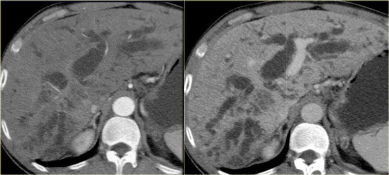
Oriental Cholangiohepatitis Radiology Case Radiopaedia Org

Oriental Cholangiohepatitis Radiology Case Radiopaedia Org

The Radiology Assistant Biliary Duct Pathology

The Radiology Assistant Biliary Duct Pathology

Sonograms Showing Variable Grades Of Intrahepatic Duct Ihd Download Scientific Diagram

Sonograms Showing Variable Grades Of Intrahepatic Duct Ihd Download Scientific Diagram

Figure 1 Image Finding An Axial Ct Image Shows A 6cm Sized Heterogeneous Lobulated Mass In The Left Lateral Segment Of The Liver A Another Ct Image Shows Bilateral Intrahepatic Duct Dilatation And Multiple Left Intrahepatic Duct Stones Measuring 3 Cm In

Figure 1 Image Finding An Axial Ct Image Shows A 6cm Sized Heterogeneous Lobulated Mass In The Left Lateral Segment Of The Liver A Another Ct Image Shows Bilateral Intrahepatic Duct Dilatation And Multiple Left Intrahepatic Duct Stones Measuring 3 Cm In

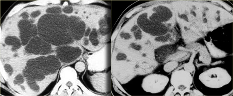
Ce Clinical Endoscopy Clin Endosc 2234 2400 2234 2443 Korean Society Of Gastrointestinal Endoscopy 10 5946 Ce 2016 150 Ce 2016 150 Case Report Diffuse Type Caroli Disease With Characteristic Central Dot Sign Complicated By Multiple Intrahepatic

Ce Clinical Endoscopy Clin Endosc 2234 2400 2234 2443 Korean Society Of Gastrointestinal Endoscopy 10 5946 Ce 2016 150 Ce 2016 150 Case Report Diffuse Type Caroli Disease With Characteristic Central Dot Sign Complicated By Multiple Intrahepatic

A Computed Tomography Ct Scan Showed Left Liver Atrophy With Ihd Download Scientific Diagram

A Computed Tomography Ct Scan Showed Left Liver Atrophy With Ihd Download Scientific Diagram

Precontrast Ct Shows Ihd And Cbd Dilatation With Several Radiopaque Download Scientific Diagram

Precontrast Ct Shows Ihd And Cbd Dilatation With Several Radiopaque Download Scientific Diagram

Source : pinterest.com